Primary sacral neurogenic tumors are rare and mostly benign tumors or low-grade malignancies. The sacrum is adjacent to vital tissues and organs, such as sacral nerves, iliac vessels, bladder, rectum, uterus, etc. Complex pelvic anatomic structure poses a great challenge to the complete resection of primary sacral tumors. Traditional open surgery can be performed via the anterior, posterior, and combined anterior-posterior approaches, with common perioperative complications including massive hemorrhage, nerve damage, and unhealed wounds. To achieve good therapeutic outcomes, the robotic system is increasingly applied in abdominal surgery because of its magnified vision, precise movement, and high flexibility. Several reports have demonstrated its efficiency in neurogenic sacral tumor resection.
One of the limitations of robotic system is that it is not designed for osteotomy and not compatible with traditional osteotomes. The ultrasonic osteotomy surgical system was useful in minimally invasive osteotomy but its application in robotic surgery has not been reported.
Objective
Aim of this study was to report the largest single-center series of robotic-assisted benign sacral neurogenic tumor resection to date, propose a new surgical strategy based on robotic-assisted benign sacral neurogenic tumor resection, and describe its application. The further objective of this study was to report for the first time the use of ultrasonic osteotomy surgical system combined with robotic system in the resection of neurogenic sacral tumors.
Patient Population
Patients who underwent primary benign sacral neurogenic tumor resection by the da Vinci Si surgical system (Intuitive Surgical Inc) at the hospital between May 2015 and March 2021 were reviewed in this study.
Surgical Strategy
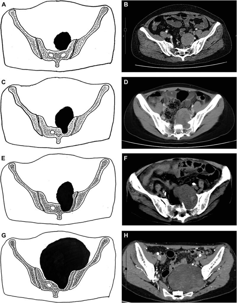
- Benign sacral neurogenic tumors was divided into 4 types based on tumor size, and the anatomic relationship between tumors and the sacrum, using preoperative imaging studies (Figure 1).
- For the presacral tumors not involving the sacrum with a maximum diameter <10 cm, they were classified as type I. For those involving the sacrum, apart from the tumor size, the tumor base in the sacrum and the number of sacral nerve root levels involved by tumors were also considered.
- Type II and III were tumors involving the sacrum with a maximum diameter <10 cm. If the maximum diameter of the tumor base in the sacrum was shorter than half of the maximum diameter of the whole tumor, it was classified as type II (narrow base). Otherwise, it was classified as type III (broad base).
- Type IV was composed of neurogenic sacral tumors involving sacral nerve roots ≥2 levels and/or with a maximum diameter ≥10 cm, with or without involving the sacrum.
- Different surgical approaches and surgical methods were applied to 4 types of benign sacral neurogenic tumors.
- Type I, II, and III neurogenic sacral tumors were resected and removed by the robotic system via an anterior approach.
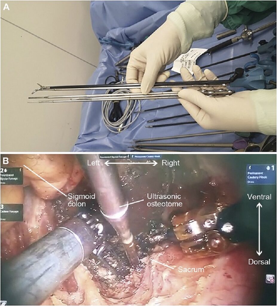
- Ultrasonic osteotomy was performed in type II and III neurogenic sacral tumors for adequate exposure of the tumors in the sacrum by the ultrasonic osteotomy surgical system (Figure 2).
- For type IV tumors, the robotic-assisted anterior approach was combined with an open posterior approach.


Figure 1. A new surgical strategy for benign sacral neurogenic tumor resection. A and B, Type I: Presacral tumors with a maximum diameter <10 cm. C and D, Type II: Tumors involving the sacrum with a maximum diameter <10 cm (narrow base). E and F, Type III: Tumors involving the sacrum with a maximum diameter <10 cm (broad base). G and H, Type IV: Tumors involving sacral nerve roots ≥2 levels with diameter ≥10 cm, with/without involving the sacrum. Adapted from source


Figure 2. A, the ultrasonic osteotome was specially designed for robotic surgery and matched robotic arms. B, the ultrasonic osteotome was used to create a bony window during robotic-assisted neurogenic sacral tumor resection. Adapted from source
Results
Clinical Characteristics and Types of Neurogenic Sacral Tumors
- Twelve patients were included in this study, including 4 male and 8 female patients. (Table)
- The median age was 48.5 years (range 22-71 years).
- The diagnosis of the patients were composed of schwannoma (9 cases), ganglioneuroma (2 cases), and neurofibroma (1 case).
- According to the surgical strategy, they were classified into type I (5 cases), type II (5 cases), type III (1 case), and type IV (1 case).
- The median tumor maximum diameter was 6.3 cm (range 4.3-8.3 cm), 5.6 cm (range 5.4-7.7 cm), 5.6 cm, and 10.3 cm for type I, type II, type III, and type IV patients, respectively.
| Case | Sex | Age (y) | Histological diagnosis | Maximum tumor diameter (cm) | Tumor base size within sacrum (cm) a | Type | Adjacent tissue | Approach | Operative time (min) | Blood loss (mL) | Ultrasonic osteotomy | Total/partial resection | Postoperative hospital stay (d) | Complication | Local recurrence/progression | Follow-up (m) |
| 1 | Female | 50 | Schwannoma | 4.4 | – | I | Iliac vessels | A | 60 | 50 | No | Total | 3 | No | No | 70.9 |
| 2 | Female | 42 | Schwannoma | 8.3 | – | I | Iliac vessels | A | 90 | 100 | No | Total | 8 | No | No | 65.3 |
| 3 | Female | 47 | Schwannoma | 4.3 | – | I | Iliac vessels, uterus | A | 70 | 100 | No | Total | 4 | Left leg muscle soreness | No | 46.2 |
| 4 | Female | 50 | Schwannoma | 5.8 | – | I | Abdominal aorta | A | 110 | 50 | No | Total | 4 | No | No | 36.7 |
| 5 | Male | 56 | Schwannoma | 8.1 | – | I | Ureter, internal iliac vessels | A | 110 | 800 | No | Total | 8 | Right foot muscle soreness | No | 17.5 |
| 6 | Male | 50 | Schwannoma | 5.5 | 1.6 | II | Abdominal aorta | A | 90 | 400 | No | Total | 4 | No | No | 51.3 |
| 7 | Female | 24 | Schwannoma | 6.5 | 2.4 | II | Ureter, internal iliac vessels | A | 80 | 30 | Yes | Total | 5 | No | No | 18.3 |
| 8 | Female | 55 | Neurofibroma | 7.7 | 2.5 | II | Branches of internal iliac vessels | A | 120 | 300 | No | Total | 4 | Left foot tingling | No | 34.1 |
| 9 | Female | 25 | Ganglioneuroma | 5.4 | 2.0 | II | Ureter, internal iliac vessels | A | 200 | 800 | No | Total | 4 | Right foot tingling | No | 14.8 |
| 10 | Male | 32 | Ganglioneuroma | 5.6 | 1.4 | II | Internal iliac vessels | A | 240 | 30 | Yes | Total | 3 | No | No | 7.9 |
| 11 | Male | 71 | Schwannoma | 5.6 | 5.2 | III | Internal iliac vessels | A | 300 | 100 | Yes | Partial | 7 | Left foot muscle soreness | No | 15.2 |
| 12 | Female | 22 | Schwannoma | 10.3 | 2.6 | IV | Intestine, internal iliac vessels, uterus | A + P | 200 | 1200 | No | Total | 5 | No | No | 22.8 |
A, anterior; P, posterior.
aMaximum diameter.
Table. The Clinical Characteristics of 12 Patients. Adapted from source
Conclusion
This new surgical strategy was helpful in guiding robotic-assisted benign sacral neurogenic tumor resection. The robotic system can be applied to benign sacral neurogenic tumor resection for type I, type II, and type III patients, and used as an adjuvant method for type IV patients. The ultrasonic osteotomy surgical system was an effective tool for type II and III patients, especially type II patients in robotic surgery.

