Introduction
Vitrectomy, or the surgical removal of vitreous, is a required step in most vitreo-retinal surgical procedures. Vitreous is a transparent gel-like substance found between the lens capsule and retina. It is composed of an inflated and highly hydrated collagen matrix with 99 wt.% water, 0.9 wt.% salts, and less than 0.1 wt.% collagen and hyaluronic acid. Vitreous is related to various functions, including maintaining transparency, protecting the retina from trauma, and other metabolic requirements. Due to the highly connected matrix of collagen fibers, vitreous removal can cause traction in areas of localized retinal adhesion. Thus, vitrectomy must temper extraction speed with the cutting action to maintain retina safety. Conventional pneumatic blade (PB) cutters use a “guillotine-style” cutter to aspirate vitreous out of the eye (Fig. 1)
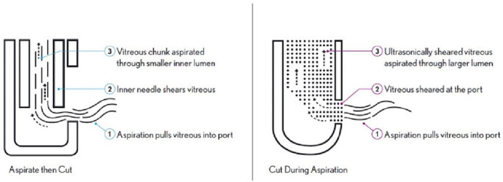

Figure 1. Comparison of pneumatic and ultrasonic mechanisms of action. (Left) Conventional pneumatic mechanisms aspirate intact vitreous and use inner needle blade to shear the tissue inside the needle lumen. (Right) Ultrasonic mechanisms shear the vitreous at port entrance, aspirating only ultrasonically sheared tissue. Figure courtesy of Bausch & Lomb. Adapted from source
Objective
The purpose of this study was to assess the performance of ultrasonic (US) vitrectomy devices by quantifying and comparing its impact on extracted vitreous properties to conventional pneumatic blade (PB) cutters using micro-extensional rheology. US vitrectomy is a new technology that offers an alternative to PB cutters used in vitreo-retinal surgeries.
Methods
Vitreous Extraction
Pig vitreous was used as a tissue analog for human vitreous, extracted from eyes using two different vitreous cutters: a PB cutter and a US cutter. Freshly harvested, unscalded, enucleated pig eyes were purchased from Sierra Medical Supplies. Preparation of each eye was performed under a surgical microscope. Eyes were acquired within 24 hours postmortem, and the tests were performed within 8 hours of receipt and 4 hours post-vitrectomy to ensure consistency in results. All eyes were stored at 4°C prior to the experiments.
Micro-Extensional Rheology
To analyze the extensional rheological properties of small samples, we adapted a simple rheometric method to apply a large strain rate to very small sample volumes.25 Sample volumes were typically between 50 and 100 microliters. Each was dispensed onto a hydrophobically treated substrate to create a domed droplet. Using a KRUSS tensiometer (K-100; KRUSS GmbH, Hamburg, Germany) in the Surface Tension setting, each sample droplet was penetrated with a 1 mm diameter platinum rod. The probe penetrated 0.5 mm into each sample. The probe was then quickly removed, so that the surface of the sample was perturbed upward. Each sample was tested independently three times. Testing was performed at room temperature, but each sample was kept at 4°C prior to testing (Fig. 2).
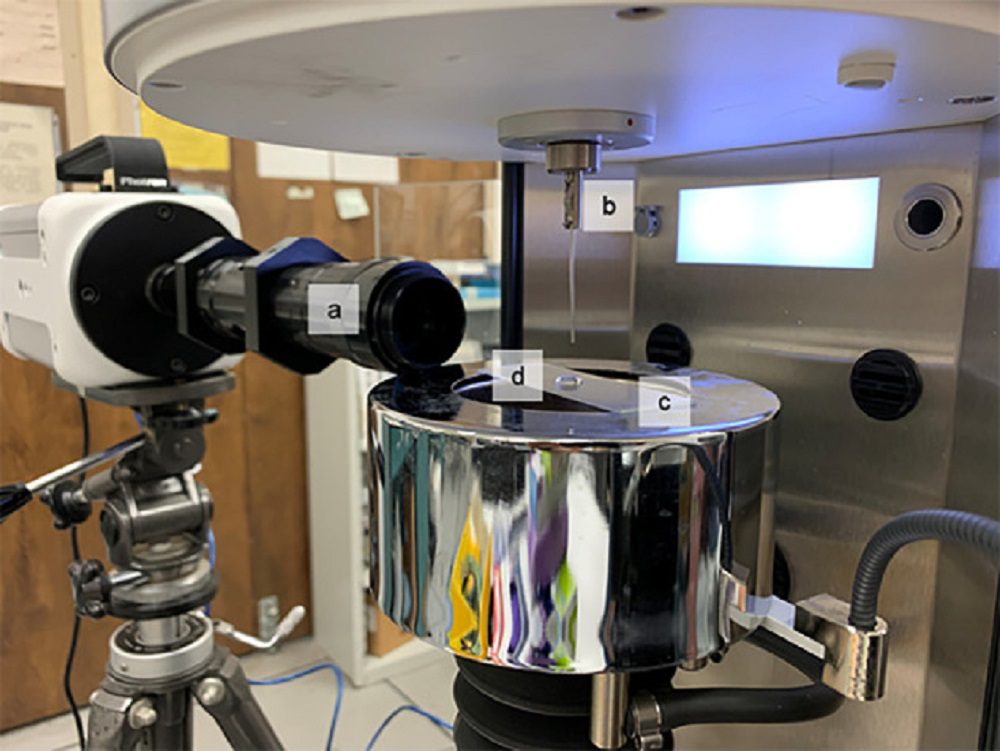

Figure 2. Micro-extensional rheology test apparatus. (a) Photron High Speed camera, (b) Kruss K-100 tensiometer with 1 mm platinum probe, (c) hydrophobically treated surface, and (d) liquid sample. Adapted from source
Results
- Thirty-six vitreous samples were successfully extracted from 20 pig eyes (2 samples per eye). Twelve samples were tested after extraction with the US cutter.
- Twenty-four samples were tested after extraction with the PB cutter (see the Table).
- The extensional rheology behavior varied dramatically between the US and PB cutter vitreous samples (Figs. 3)
- Extensional relaxation times (Fig. 4) averaged 27.25 ms for the PB samples, and 0.37 ms for the US samples, a reduction of approximately 99%.
- The average extensional relaxation time for the US samples was significantly lower than the PB samples (P < 0.001) with up to two orders of magnitude in difference.
- The inertia-capillary regime of the US samples dominated the flow, and the elasto-capillary liquid bridge existed for only a few image frames in each trial, indicating the protein interactions had little effect on the flow in these samples.
- The relatively high extensional relaxation time for PB samples culminating in beads-on-a-string structures indicate the elasto-capillary effects of the protein interactions were significant. This is an indication of relatively large protein fragments and high cross-linking of the proteins within the samples.
Summary of Cutter Configuration and Resultant Extensional Relaxation Times in Milliseconds
| Vitrectomy Method | λE (ms) | ||||
| Number of Samples | Cutter Size (Gauge) | Vacuum Pressure (mm Hg) | Mean | Stdev | |
| Ultrasonic | 3 | 23 | 100 | 0.17 | 0.10 |
| 3 | 23 | 300 | 0.39 | 0.10 | |
| 3 | 25 | 100 | 0.13 | 0.06 | |
| 3 | 25 | 300 | 2.34 | 2.46 | |
| (Total) | (12) | (0.37) | (1.50) | ||
| Pneumatic blade | 4 | 23 | 100 | 25.79 | 12.95 |
| 4 | 23 | 300 | 24.17 | 15.33 | |
| 4 | 23 | 600 | 37.63 | 21.17 | |
| 4 | 25 | 100 | 19.11 | 8.47 | |
| 4 | 25 | 300 | 23.47 | 12.41 | |
| 4 | 25 | 600 | 33.33 | 11.56 | |
| (Total) | (24) | (27.25) | (15.08) | ||
Stdev, standard deviation.
Table. Summary of Cutter Configuration and Resultant Extensional Relaxation Times in Milliseconds. Adapted from source
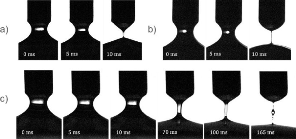

Figure 3. Radius evolution images starting at approximately 60% radius, taken 5 ms apart. (a) Balanced saline solution, containing no polymer or protein and showing no extensional relaxation behavior. (b) Vitesse ultrasonic cutter vitreous. (c) BiBlade pneumatic cutter vitreous at longer time steps. Beads-on-a-string phenomenon was observed at the end of the liquid bridge evolution (at 165 ms). Adapted from source
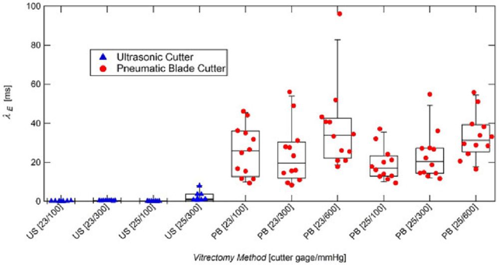

Figure 4. Box plots of sample relaxation times extracted using ultrasonic and pneumatic blade cutters. The average extensional relaxation time for US and PB samples were 0.37 ms and 27.25 ms, respectively. Adapted from source
Conclusion
In summary, ultrasonic vitrectomy technology was assessed and its performance compared to conventional PB vitrectors. By using extensional rheology, it was showed that significant differences in the vitreous samples extracted using the two cutter styles, where typical shear rheology was not likely to do so. When extracted using an ultrasonic cutter, the extensional relaxation times of the vitreous samples were orders of magnitude shorter compared to samples extracted using the pneumatic blade cutter (US sample mean = 0.37 ms, and PB sample mean = 27.25 ms). These results confirm that the US cutter breaks the vitreous collagen into very small fragments compared to pneumatic cutters. This has broad implications for improved ergonomics and safety of the ultrasonic cutter, as well as for the development and improvement of vitrectomy devices and techniques using US technology.

